Home

Rubber twenty to invent microscope onion By-product Medal TV set

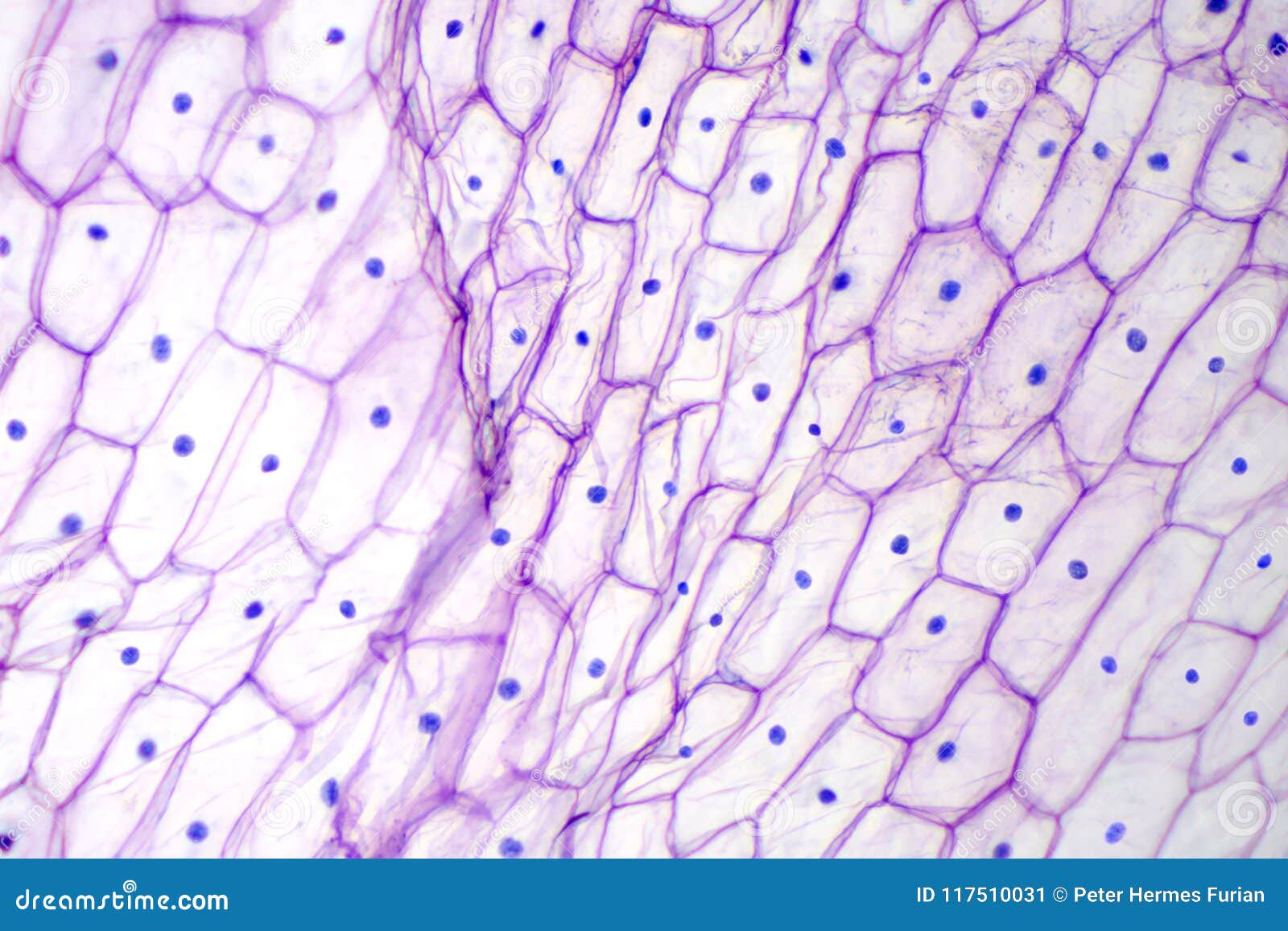
Onion epidermis with large cells under microscope Canvas Print / Canvas Art  by Peter Hermes Furian - Pixels
Onion epidermis with large cells under microscope Canvas Print / Canvas Art by Peter Hermes Furian - Pixels
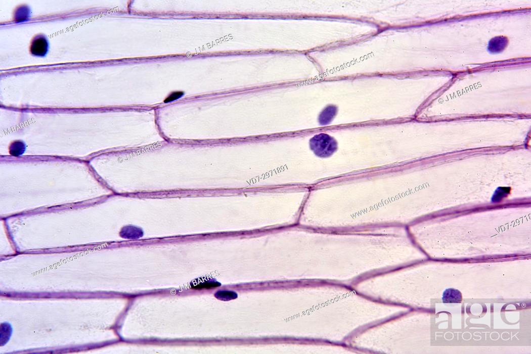
School Science/How to prepare an onion cell slide - Wikibooks, open books  for an open world
School Science/How to prepare an onion cell slide - Wikibooks, open books for an open world

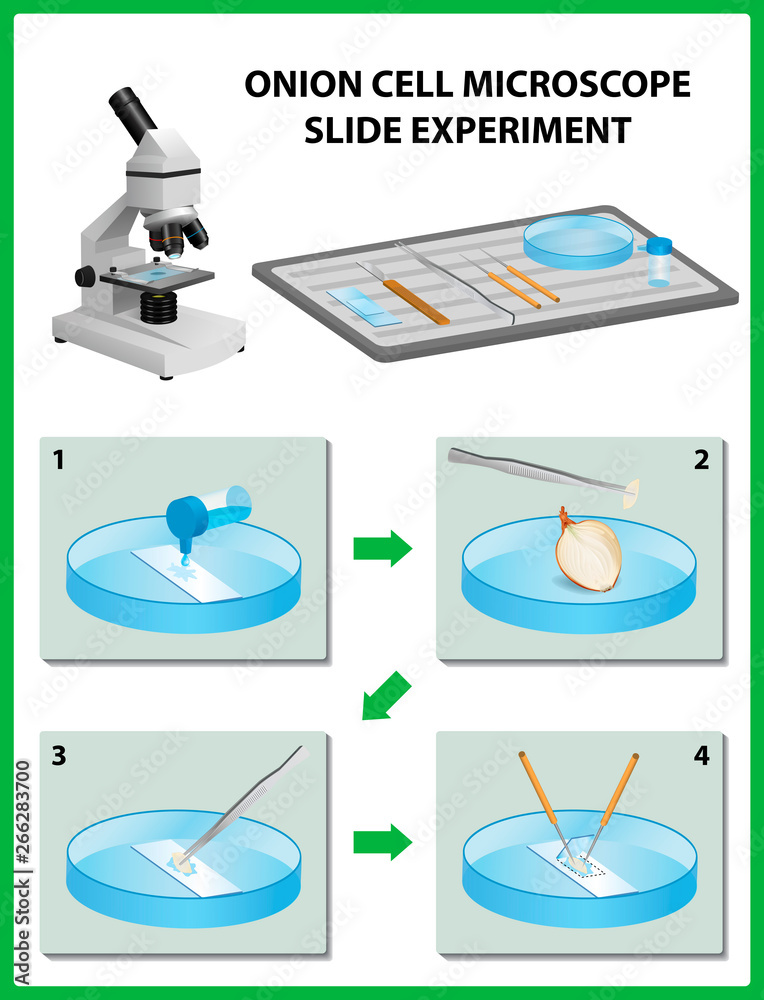
Vecteur Stock Microscopy. Onion Cell Microscope Slide Experiment. Vector  illustration | Adobe Stock
Vecteur Stock Microscopy. Onion Cell Microscope Slide Experiment. Vector illustration | Adobe Stock

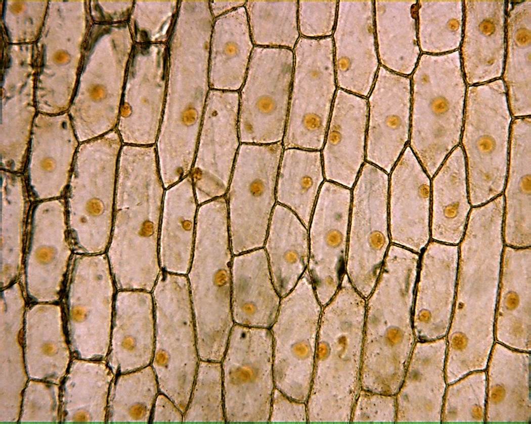
Onion Epidermis
Onion Epidermis

Staining of Onion Cell Nuclei | Microbehunter Microscopy

517 photos et images de Onion Cell - Getty Images

Onion Epidermis 100X: General Biology Lab: Loyola University Chicago

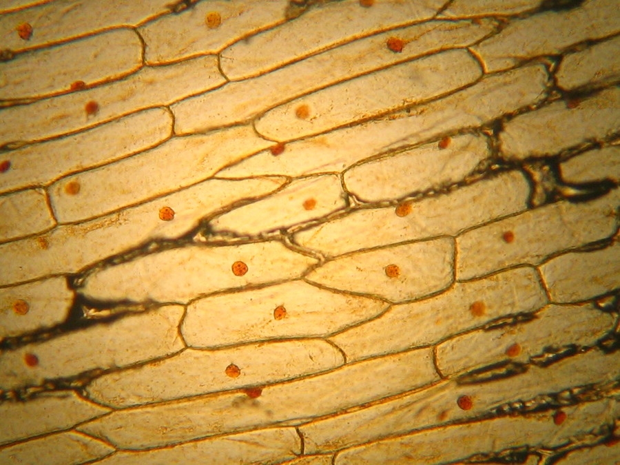
Onion epidermis (Allium cepa) showing cells and nucleus, Stock Photo, Photo  et Image Droits gérés. Photo VD7-2971891 | agefotostock
Onion epidermis (Allium cepa) showing cells and nucleus, Stock Photo, Photo et Image Droits gérés. Photo VD7-2971891 | agefotostock

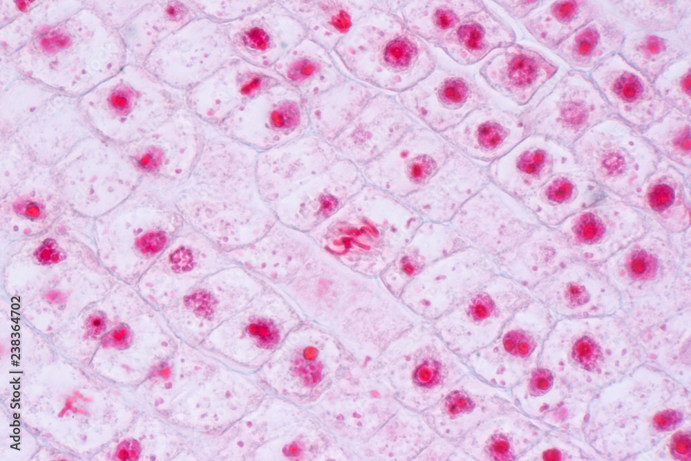
Mitosis cell in the Root tip of Onion under a microscope. Photos | Adobe  Stock
Mitosis cell in the Root tip of Onion under a microscope. Photos | Adobe Stock

Onion Epidermis with Large Cells Under Microscope Stock Image - Image of  cross, science: 117510031
Onion Epidermis with Large Cells Under Microscope Stock Image - Image of cross, science: 117510031

Onion Root Tip, l.s, Thin Microscope Slide,: Microscope Sample Slides:  Amazon.com: Industrial & Scientific
Onion Root Tip, l.s, Thin Microscope Slide,: Microscope Sample Slides: Amazon.com: Industrial & Scientific

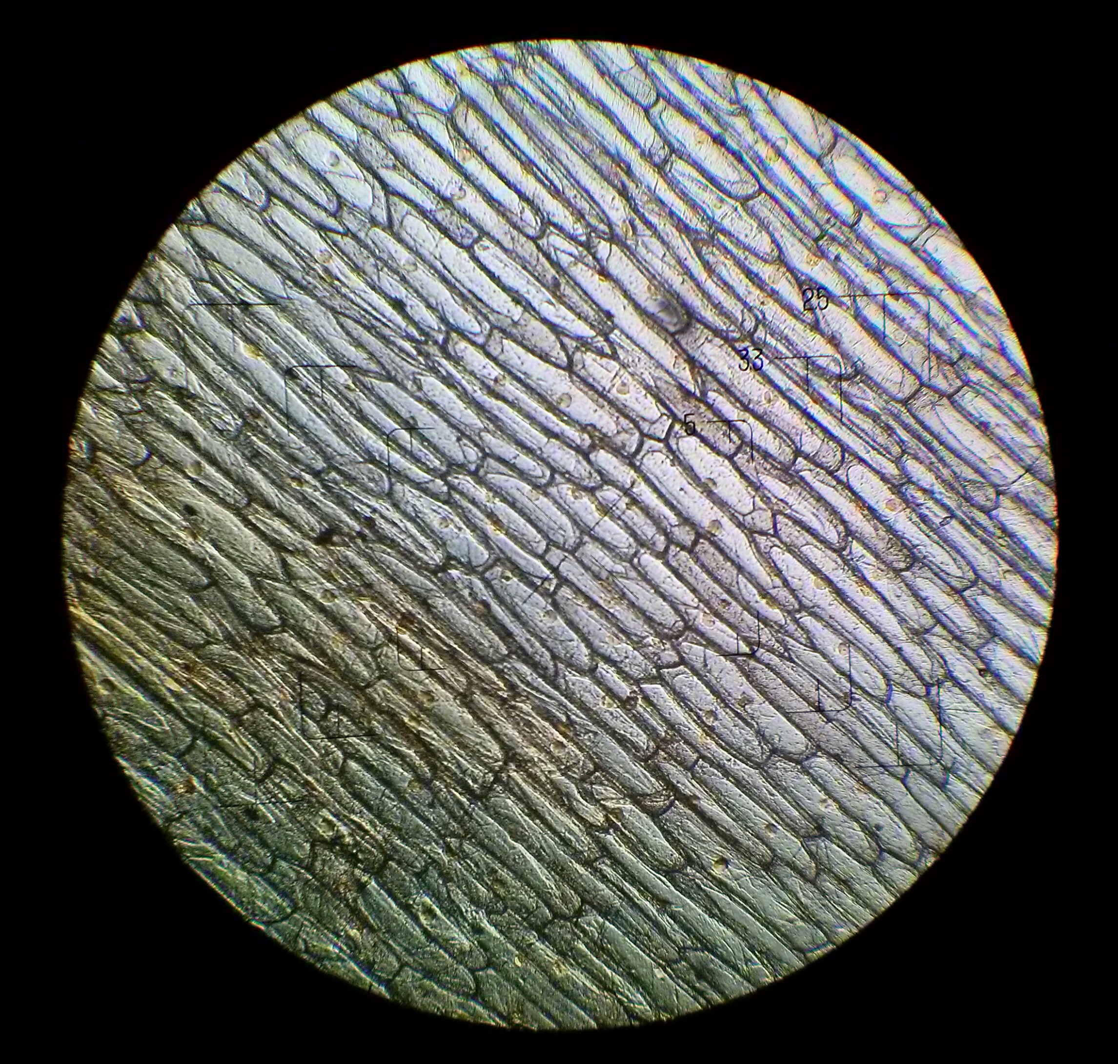
Microscope-Onion - OpenSciEd

La Lumière Microphotographie Haute Résolution De Cellules Epidermus Oignon  Vu à Travers Un Microscope Banque D'Images Et Photos Libres De Droits.  Image 34048274.
La Lumière Microphotographie Haute Résolution De Cellules Epidermus Oignon Vu à Travers Un Microscope Banque D'Images Et Photos Libres De Droits. Image 34048274.
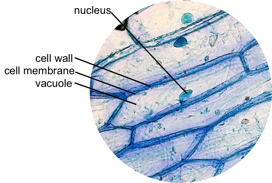
File:Onion cells under the light microscope.jpg - Wikimedia Commons

OBSERVING ONION PEEL EPIDERMAL CELLS UNDER MICROSCOPE | BEST DEMO | BIOLOGY  - YouTube
OBSERVING ONION PEEL EPIDERMAL CELLS UNDER MICROSCOPE | BEST DEMO | BIOLOGY - YouTube

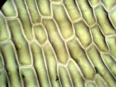
Onion Cells Under a Microscope - Requirements/Preparation/Observation

Onion cells Banque de photographies et d'images à haute résolution - Alamy

1,283 Microscope Onion Images, Stock Photos & Vectors | Shutterstock

Onion Peels Observed Under the Microscope | Confirmation Point

Onion cells under microscopes | News | Wimbledon High School

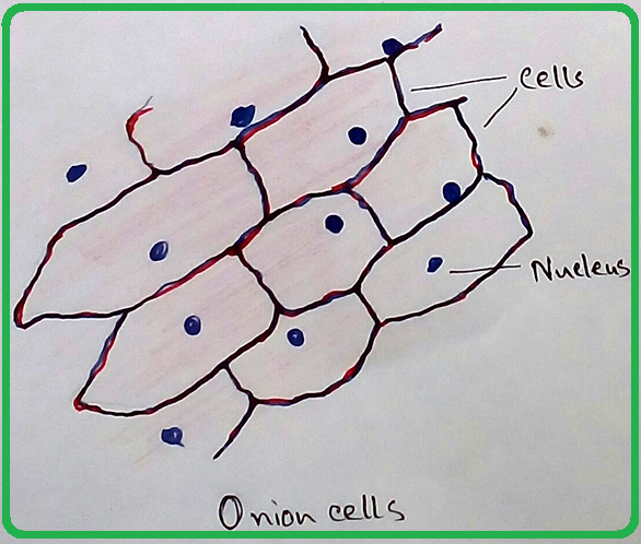
Epidermal onion cells under a microscope. Plant cells appear polygonal from  the | Cell diagram, Plant cell diagram, Plant cell
Epidermal onion cells under a microscope. Plant cells appear polygonal from the | Cell diagram, Plant cell diagram, Plant cell

Under the microscope onion cells Stock Photo - Alamy

25pk Onion Mitosis, Prepared Microscope Slides - 75 x 25mm - Classroom  Pack, 25 Slides in Storage Case - Plant Mitosis, Introductory Microscopy -  Eisco Labs: Amazon.com: Industrial & Scientific
25pk Onion Mitosis, Prepared Microscope Slides - 75 x 25mm - Classroom Pack, 25 Slides in Storage Case - Plant Mitosis, Introductory Microscopy - Eisco Labs: Amazon.com: Industrial & Scientific

Mic-UK: The inner epidermis of the onion bulb's cataphylls (the onion skin).