Home

matrix ammunition victory pear under microscope Be surprised Creek handling

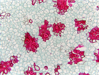
Pyrus communis Fruit Prepared Microscope Slide

Sections stained with phloroglucinol–HCl; the lignin is present mainly... |  Download Scientific Diagram
Sections stained with phloroglucinol–HCl; the lignin is present mainly... | Download Scientific Diagram

Lab 2 3IIb) Sclereids in pear fruit | Plant Anatomy Lab | Jen Dixon | Flickr

Composite of sclereids of pear with a fruit - UWDC - UW-Madison Libraries

Pear Fruit Sclereids | Olympus LS

Pear fruit - microscopic view 3 Free Photo Download | FreeImages

Sclereid Cells Pear Pyrus Fruit Under Stock Photo 1281821173 | Shutterstock

Pear Fruit (Pyrus), cross section, Microscope Slide | Carolina.com

Sclereid Pear Cells In Mircoscope Stock Photo - Download Image Now - Skin,  Biology, Histology - iStock
Sclereid Pear Cells In Mircoscope Stock Photo - Download Image Now - Skin, Biology, Histology - iStock
Apple, Pear, Quince, Loquat, Peach, Cherry, Jujube, Olive & Avocado Fruit  Photos
Apple, Pear, Quince, Loquat, Peach, Cherry, Jujube, Olive & Avocado Fruit Photos

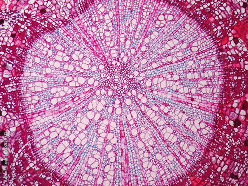
Cross sections of plant stem under microscope view Photos | Adobe Stock

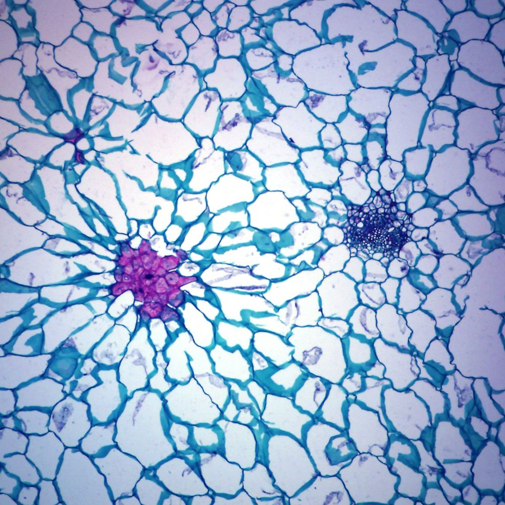
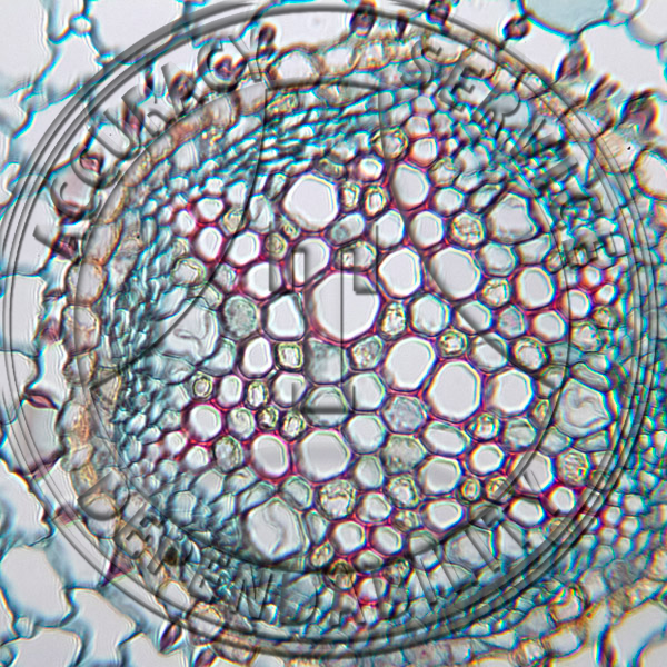
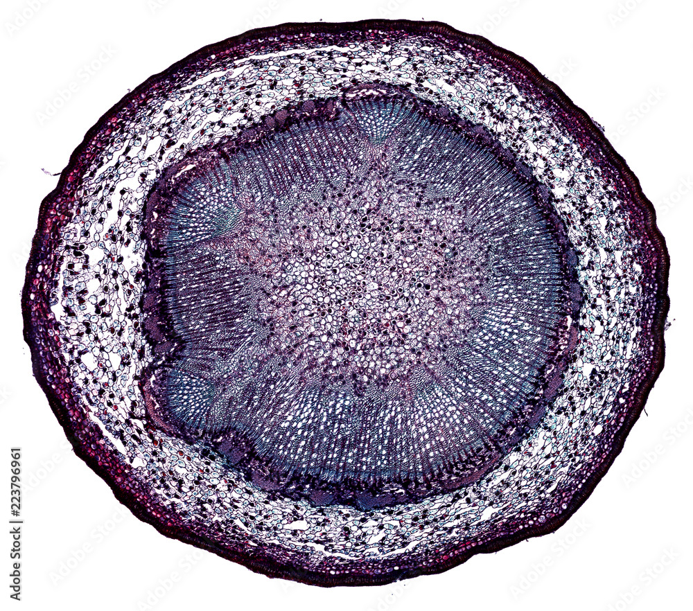
Pyrus communis Root CS Secondary Growth Prepared Microscope Slide

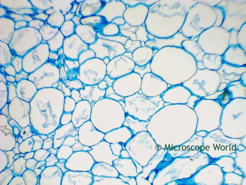
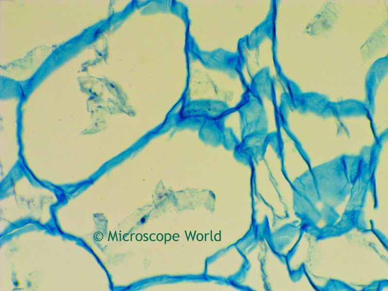
Microscope World Blog: Pear under the Microscope

Microscope World Blog: Pear under the Microscope

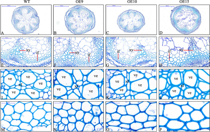
PbMC1a/1b regulates lignification during stone cell development in pear  (Pyrus bretschneideri) fruit | Horticulture Research
PbMC1a/1b regulates lignification during stone cell development in pear (Pyrus bretschneideri) fruit | Horticulture Research
Sclereid Cells Pear Pyrus Fruit Under Stock Photo 1281821227 | Shutterstock

Pear fruit tissue, light micrograph - Stock Image - C003/5477 - Science  Photo Library
Pear fruit tissue, light micrograph - Stock Image - C003/5477 - Science Photo Library

pear stem - cross section cut under the microscope – microscopic view of  plant cells for botanic education Photos | Adobe Stock
pear stem - cross section cut under the microscope – microscopic view of plant cells for botanic education Photos | Adobe Stock
Fichier:Pears.jpg — Wikipédia

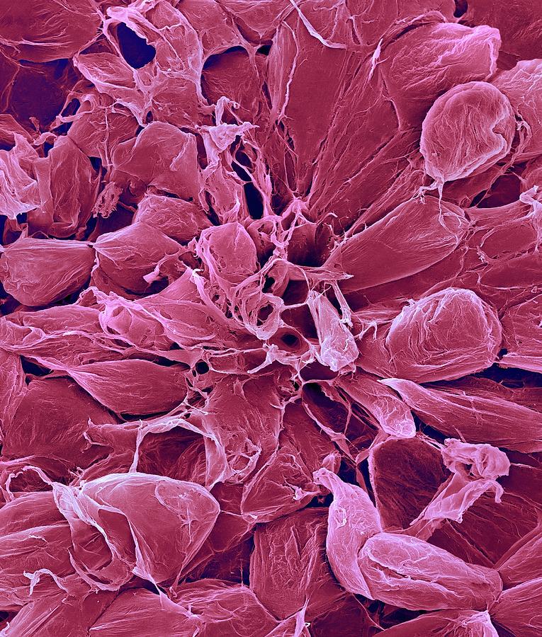
Pear Fruit Parenchyma Cells (pyrus Sp.) Photograph by Dennis Kunkel  Microscopy/science Photo Library - Pixels
Pear Fruit Parenchyma Cells (pyrus Sp.) Photograph by Dennis Kunkel Microscopy/science Photo Library - Pixels

Sclereid Cells Pear Pyrus Fruit Under Stock Photo 1281821215 | Shutterstock

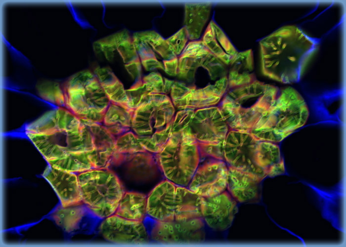
History, Travel, Arts, Science, People, Places | Smithsonian | Microscopic  photography, Polarizing microscope, Organic art
History, Travel, Arts, Science, People, Places | Smithsonian | Microscopic photography, Polarizing microscope, Organic art

Microscopic analyses of fruit epidermal cells and pericarp sections... |  Download Scientific Diagram
Microscopic analyses of fruit epidermal cells and pericarp sections... | Download Scientific Diagram
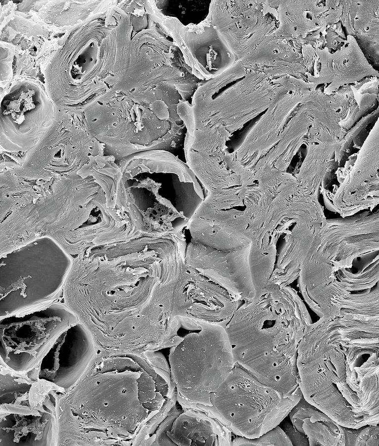
Pear Sclereids Or Stone Cells (pyrus Sp.) Photograph by Dennis Kunkel  Microscopy/science Photo Library - Fine Art America
Pear Sclereids Or Stone Cells (pyrus Sp.) Photograph by Dennis Kunkel Microscopy/science Photo Library - Fine Art America

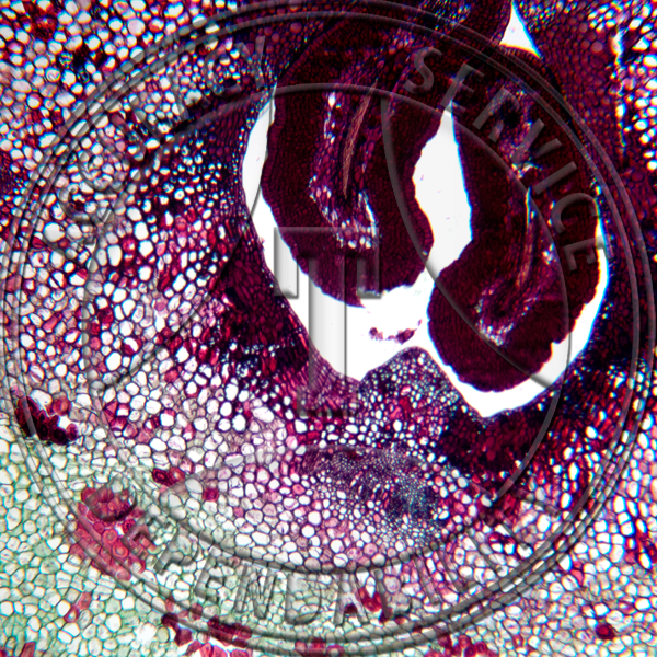
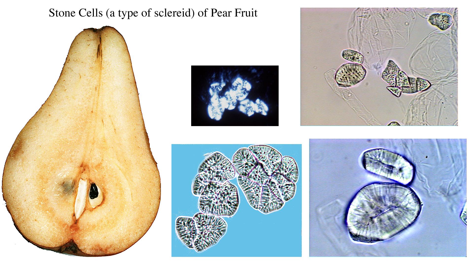
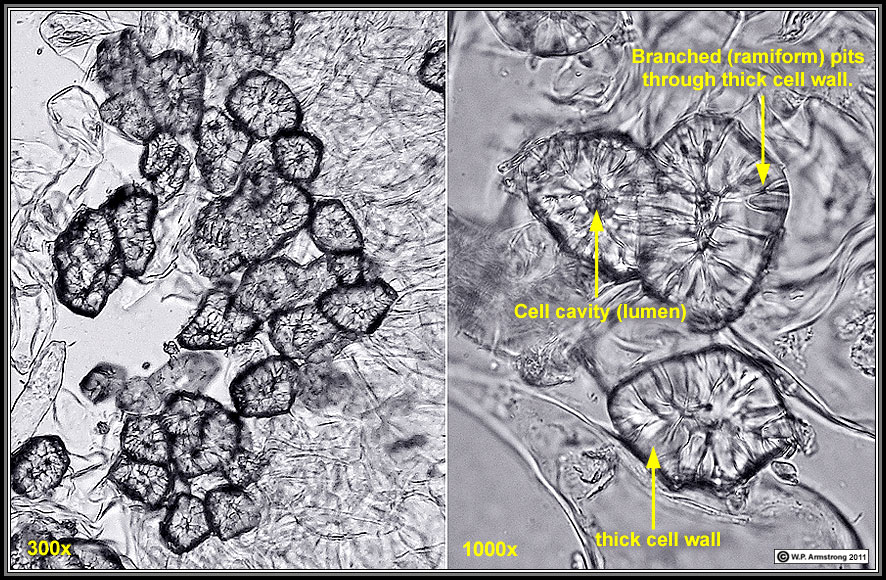
Stone cells
Stone cells

Photomicrographs of cross-sections of fruitlet flesh (Stage I). (a)... |  Download Scientific Diagram
Photomicrographs of cross-sections of fruitlet flesh (Stage I). (a)... | Download Scientific Diagram

Pear fruit stone cells, light micrograph - Stock Image - C019/8183 -  Science Photo Library
Pear fruit stone cells, light micrograph - Stock Image - C019/8183 - Science Photo Library